- Visibility 259 Views
- Downloads 18 Downloads
- DOI 10.18231/j.jds.2020.003
-
CrossMark
- Citation
Variations in mandibular coronoid process-A morphometric treatise
- Author Details:
-
Sukhman Kahlon
-
Gaurav Agnihotri *
Introduction
Coronoid process in Greek means “like a crown”. In lower animals, separate coronoid bones are present which articulate with the splenial, angular and supra angular bones to form a common “dentary bone” which is homologous to mandible in humans.[1]
The coronoid process of mandible is of clinical significance for maxillofacial surgeons for reconstructive purposes as it is used as graft for reconstruction of osseous defects in oral and facio-maxillary region.[2] Anatomical variations in the coronoid process can result in extremely narrow vestibular space due the close proximity of the medial aspect of the coronoid process to the distal molar.[3]
The present morphometric treatise focuses on variant anatomy of the coronoid process in Indian population.The study provides baseline data for Indian population which has clinical implications and repercussions.
Materials and Methods
The material for the present study comprised of 500 adult human mandibles. These mandibles were obtained from the different medical colleges in the state of Punjab. Any mandible broken or dysmorphic were excluded from the study.
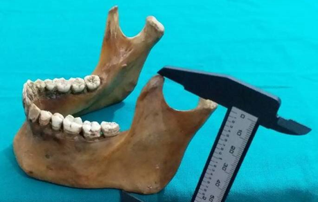
The coronoid height was measured as distance between the coronoid and most protruding portion of inferior border of the ramus of mandible ([Figure 1]).
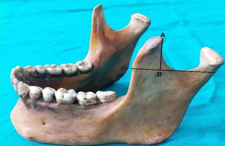
The length of coronoid process was taken from the line tangential to the deepest part of mandibular notch to apex of the coronoid process ([Figure 2]).
Results
The shape of coronoid process was classified into 3 types: ([Figure 3])
Triangular: Tip pointing directly upwards.
Rounded: Tip rounded.
Hook: Tip pointing backwards.
Three incidence percentage for round, triangular and hook shaped coronoid processes came out to be 46, 42 and 12 respectively. The mean value of height and length of coronoid process was determined to be 60.62 mm and 12.53 mm respectively. ([Table 1], [Table 2] )
Discussion
The coronoid process develops as a discrete entity within the mass of the temporalis muscle anlage, subsequently it unites with the main portion of mandibular ramus at approximately eight weeks of age.[4] The largest portion of temporalis muscle is attached to the apex, whole of the medial surface and anterior part of lateral surface of coronoid process. The remaining part of lateral surface provides attachment to anterior fibres of masseter. These two are important muscles of mastication which show morpho functional dependence.
Several authors have described the various shapes of coronoid process. According to which, some processes are triangular, hook shaped and rounded [5], [6], [7] whereas others have described it as beak shaped.[8] The shape and size of coronoid process is influenced by dietary habit, genetic constitution, hormonal and mainly by temporalis muscle activity.[9] In the present study, the incidence of the rounded shape was the highest followed by the triangular shape. In most studies in India the triangular shape was most commonly observed. In previous studies in Turkish population[10] and Bangladeshi population[11] the most common shape observed was the hook shape. ([Table 3], [Table 4])
The coronoid process projects upwards and slightly forwards. It has a top border and is convex in shape, while its lower part is concave in shape. Its margins and medial surface provide attachment to temporalis muscle. The coronoid process is suitable for paranasal augmentation. Its clinical application is also favourable because its size and morphology fits into the paranasal region, with the additional advantages of biocompatibility, availability, and reduced operation time for harvesting.[12], [13] The present study focuses on the height and length of coronoid process of mandible with the aim to characterize the morphological profile of the coronoid process in Indian population. This will benefit dental surgery, anthropological and forensic practice.
Coronoid process enlargement may be seen in some pathological conditions like osteochondroma, exostosis, osteoma and other developmental anomalies. Though fracture of mandible is common, but incidence of coronoid fracture is rare (2%) and requires no treatment unless impingement on the zygomatic arch is present. Coronoid process hyperplasia is a very rare cause of mandibular hypomobility. So, it is usually underdiagnosed, but a thorough background anatomical knowledge can help in examining the patient clinically and radiologically.[14], [15] This ultimately will help in the line of management and a better clinical outcome.[16]
The coronoid process can be removed intra-orally without any functional deficiency and facial disfigurement. It is expected that a knowledge of the morphometric profile of the coronoid process in different populations will aid the clinician in reconstruction procedures such as those pertaining to orbit floor, alveolar defects, paranasal sinus augumentation, non union fracture mandible, osseous defects reconstruction and other repair procedures in cranio-maxillo facial surgeries.[17], [18]


|
Shape |
Number of mandibles |
Percentage incidence |
|
Triangular |
210 |
42% |
|
Hook Shaped |
060 |
12% |
|
Rounded |
230 |
46% |
|
Parameters |
Mean value |
Standard deviation |
|
Height |
60.62 |
5.95 |
|
Length |
12.53 |
2.75 |
|
Authors (Year) |
Population |
Triangular |
Hook shaped |
Rounded |
|
Khan TA and Sharieff JH [5] (2011) |
South India |
67% |
30% |
3% |
|
Prajapati VP et al [6](2011) |
Western Indians |
54.17% |
21.25% |
24.58% |
|
Desai VC et al [7](2014) |
South-west Indians |
136 (68%) |
48 (24%) |
16 (8%) |
|
Pradhan S et al [1] (2014) |
Eastern Indians |
86 (46.73%) |
33 (17.93%) |
65 (35.3%) |
|
Present study (2020) |
Indians |
210 (42%) |
60 (12%) |
230 (46%) |
|
Authors (Year) |
Population |
Mean ± SD |
|
Koyama M [12] (1965) |
Japanese |
98.27±5.2 (male) 91.85±5.6 (female) |
|
Kumar MP and Lokanadham S [13] (2013) |
South-east Indians |
59.37±5.03 |
|
Sandeepa NC et al [14] (2017) |
Saudi |
74.18±5.78 (male) 63.28±5.06 (female) |
|
Present study (2020) |
Indians |
60.62±5.95 |
Conclusions
Our study of coronoid process suggest that round shape is the most common presentation(46%) followed by triangular(42%) and then hook shaped(12%). This morphometric treatise provides valuable inputs relevant for anthropological comparisons, forensic investigations and reconstructive procedures.
Source of Funding
None.
Conflict of Interest
None.
References
- S S Pradhan, D Bara, S Patra, S Nayak, C Mohapatra. Anatomical Study of Various Shapes of Mandibular Coronoid Process in Relation to Gender & Age. IOSR J Dent Med Sci 2014. [Google Scholar]
- P H Choung, S G Kim. The coronoid process for paranasal augmentation in the correction of midfacial concavity. Oral Surg Oral Med Oral Pathol Oral Radiol Endod 2001. [Google Scholar]
- S M Mintz, A Ettinger, T Schmakel, M J Gleason. Contralateral coronoid process bone grafts for orbital floor reconstruction: An anatomic and clinical study. J Oral Maxillofac Surg 1998. [Google Scholar]
- M N Spyropoulos. The morphogenetic relationship of the temporal muscle to the coronoid process in human embryos and fetuses. Am J Anat 1977. [Google Scholar]
- T A Khan, J H Sharieff. Observation on morphological features of human mandibles in 200 South Indian subjects. Anatomical Karnataka 2011. [Google Scholar]
- V P Prajapati, O Malukar, S K Nagar. Variations in the morphological appearance of the coronoid process of human mandible. National J Med Res 2011. [Google Scholar]
- V C Desai, S D Desai, H S Shaik. Morphological study of mandible. J Pharm Sci Res 2014. [Google Scholar]
- E A Schafer, G D Thane. . Quain’s Elements of Anatomy: The bones of the head 1890. [Google Scholar]
- H Kausar, A Tripathi, S Raizaday, S Bharti, S Jain, S Khare. Morphology and Morphometry of Coronoid Process of Dry Mandible- A Comprehensive Study. Journal of Evidence Based Medicine and Healthcare 2020. [Google Scholar]
- S Bakirci, Ilknur Ari, I M Kafa. Morphometric characteristics and typology of coronoid process of the mandible. Acta Med Mediterr 2013. [Google Scholar]
- A Hossain, S M, M Hossain, S M Banna, Famh. Variations in the shape of the coronoid process in the adult human mandible. Bangladesh J Anat 2011. [Google Scholar]
- M KOYAMA. An Anthropological Study on the Mandible in the Japanese, especially Measurements in Relation to the Construction of an Articulator. J Nihon Univ Sch Dent 1965. [Google Scholar]
- M P Kumar, S Lokanadham. Sex determination & morphometric parameters of human mandible. Int J Res Med Sci 2013. [Google Scholar]
- N C Sandeepa, A A Ganem, W A Alqhtani, Y M Mousa, E K Abdullah, A H Alkhayri. Mandibular indices for gender prediction: A retrospective radiographic study in Saudi population. J Dent Oral Health 2017. [Google Scholar]
- S Nayak, S Patra, G Singh, C Mohapatra, S Rath. Study of the size of the coronoid process of mandible. J Anatomical Soc India 2016. [Google Scholar]
- S Nayak, S Pradhan, D P Bara, S Patra. Bilateral elongated coronoid process: A case report. IOSR J Dent Med Sci 2015. [Google Scholar]
- S Amrani, G E Anastassov, A H Montazem. Mandibular Ramus/Coronoid Process Grafts in Maxillofacial Reconstructive Surgery. J Oral Maxillofac Surg 2010. [Google Scholar]
- L Clauser, C Curioni, S Spanio. The use of the temporalis muscle flap in facial and craniofacial reconstructive surgery. A review of 182 cases. J Craniomaxillofac Surg 1995. [Google Scholar]
How to Cite This Article
Vancouver
Kahlon S, Agnihotri G. Variations in mandibular coronoid process-A morphometric treatise [Internet]. J Dent Spec. 2020 [cited 2025 Sep 10];8(1):9-12. Available from: https://doi.org/10.18231/j.jds.2020.003
APA
Kahlon, S., Agnihotri, G. (2020). Variations in mandibular coronoid process-A morphometric treatise. J Dent Spec, 8(1), 9-12. https://doi.org/10.18231/j.jds.2020.003
MLA
Kahlon, Sukhman, Agnihotri, Gaurav. "Variations in mandibular coronoid process-A morphometric treatise." J Dent Spec, vol. 8, no. 1, 2020, pp. 9-12. https://doi.org/10.18231/j.jds.2020.003
Chicago
Kahlon, S., Agnihotri, G.. "Variations in mandibular coronoid process-A morphometric treatise." J Dent Spec 8, no. 1 (2020): 9-12. https://doi.org/10.18231/j.jds.2020.003
